
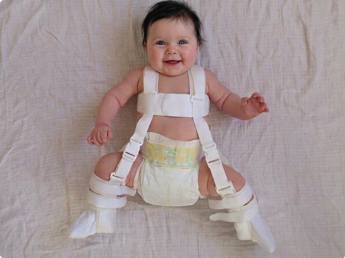
Developmental dysplasia of the hip is a spectrum, not a single condition. It ranges from a socket that is merely shallow to a femoral head that sits completely outside the joint. The Pavlik harness treats the whole spectrum in infants through one principle: hold the joint in the position where it is most stable, and let growth do the rest.
The anatomy in plain terms
The hip is a ball-and-socket joint. The ball is the femoral head at the top of the thigh bone; the socket is the acetabulum in the pelvis. At birth both are largely cartilage and both are still forming.
The socket's depth is not fixed at birth — it is shaped by the ball pressing into it. This is why a dislocated hip left untreated grows progressively shallower: without the ball inside, the socket has no template to grow around. Every week matters.
Why flexion and abduction work
When the hip is flexed above 90 degrees and gently abducted, three things happen at once. The femoral head slides back into the socket. The surrounding capsule and the psoas and adductor muscles relax rather than pulling the head out. And the direction of force through the joint points straight into the deepest part of the socket.
Hold that position continuously and the socket remodels. Ultrasound typically shows measurable improvement in the alpha angle within two to three weeks of consistent wear.
The safe zone
Clinicians talk about a safe zone: the range of abduction in which the hip stays reduced but the blood supply to the femoral head is not compromised. Too little abduction and the hip stays out. Too much — pushing the knees toward the mattress — squeezes the medial circumflex artery and risks avascular necrosis, the most serious complication of over-tight harness treatment.
This is precisely why the straps are set by a clinician and rechecked at every visit. The safe zone changes as the baby grows.
What the follow-up looks like
- Week 1: fit check, strap adjustment, skin inspection, ultrasound to confirm the hip is reduced in the harness.
- Weeks 2–6: review every one to two weeks; straps let out as the baby grows.
- Weeks 6–12: ultrasound to document socket improvement; weaning begins if the hip is stable and the alpha angle is normalising.
- After weaning: an X-ray around 6 and 12 months, and often again at walking age, to confirm the socket has matured.
When the harness is not working
If a hip is not reduced on ultrasound after two to three weeks of correct full-time wear, most surgeons stop the harness rather than persist. Continuing to hold an unreduced hip flexed against the back of the socket can erode it — a complication called Pavlik harness disease. The next step is usually an examination under anaesthetic with closed reduction and a spica cast.
Frequently asked questions
Will my baby's hips be normal afterwards?
In the large majority of infants treated early and successfully, the hip develops normally and there are no long-term restrictions on walking, running or sport. Long-term follow-up X-rays confirm this.
Can hip dysplasia come back after treatment?
Residual dysplasia occurs in a minority of children, which is why follow-up imaging continues until walking age even after a successful harness course.